IL-17A Disrupts Fetal Brain Development via Maternal Immune Activation
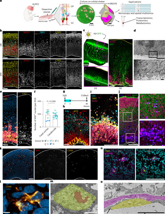
Maternal immune activation (MIA) has been shown to disrupt brain development and increase the risk of neurodevelopmental disorders (NDDs) in offspring. A recent study introduces a three-dimensional ex vivo culture system called 'cerebroids,' derived from human fetal brain tissue, which maintains the critical developmental processes and cellular diversity of the human cortex. This innovative model allows researchers to investigate the effects of IL-17A, a cytokine associated with MIA and NDDs, on cortical development. The study demonstrates that IL-17A leads to premature cortical folding, increased cortical thickness, and accelerated neurogenesis and neuronal maturation. It also reveals that IL-17A significantly disrupts extracellular matrix-related pathways and activates NF-κB signaling in neural stem cells, resulting in sustained inflammatory responses that contribute to these developmental abnormalities. Importantly, treatment with the NF-κB pathway inhibitor parthenolide was shown to reverse these effects, providing insights into potential therapeutic avenues. The research highlights the importance of understanding how maternal immune responses can influence fetal brain development and the mechanisms underlying the increased risk of neurodevelopmental disorders. The cerebroid model offers a promising platform for further exploration of these critical processes, preserving the structural integrity and cellular architecture necessary for studying both healthy neurodevelopment and the impact of environmental factors such as maternal immune activation.